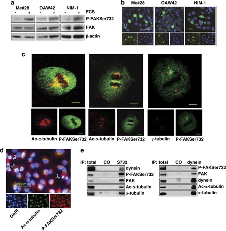
Figure 3.
P-FAKSer732 accumulates in mitotic cells and co-localizes with MTs of the mitotic spindle. (a) Western blotting on total lysates from starved or FCS-stimulated cells. (b) IF performed on fixed cells upon FCS stimulation with anti-P-FAKSer732 Ab (green). Nuclei were stained with DAPI (blue). (c) Confocal IF performed on fixed Me#28 cells with anti-P-FAKSer732 and ac-α- (left and middle panels) or γ-tubulin Abs (right panel). Images of metaphases (left and right panels, section 14 and 12, respectively) and late anaphase (section 11, middle panel) are shown. Bars, 10 μm. (d) IF performed on Me#28 cells upon extraction of free tubulin with PEM buffer (see Materials and Methods). The white arrows indicate the mitotic phases: 1, prophase; 2 anaphase; 3, telophase; 4, cytokinesis. The immunostaining was performed with anti-P-FAKSer732 (red) and ac-α-tubulin (green). Nuclei were stained with DAPI (blue). Bars, 60 μm. (e) IP on lysates from Me#28 cells with anti-P-FAKSer732 (S732) and -dynein intermediate chain Abs. Immunoprecipitated fractions were analyzed by western blotting. Normal rabbit or mouse sera were used as negative control (CO). For immunoblottings, Abs are reported on the right

