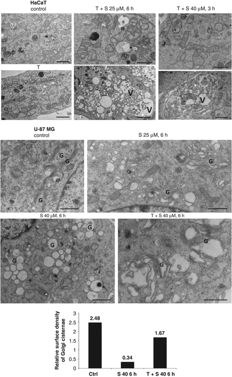
Figure 5.
Ultrastructure of siramesine-treated HaCaT and U-87MG cells by TEM. Following the treatment, cells were fixed with 1% glutaraldehyde and embedded in epon. Thin sections were analysed with TEM. The presence of Golgi cisternae in U-87MG cells in thin sections was quantified using stereology. Bars present ratios between the total length of the Golgi cisternal membrane (estimated by intersection counting) and the total cytoplasmic area (estimated by point counting). Bar size, 1 μm. V, vacuole with cytoplasmic inclusions; G, area with Golgi cisternae and vesicles; S, siramesine; T, α-tocopherol

