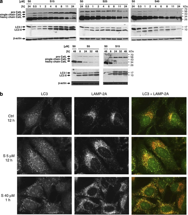
Figure 7.
Siramesine-induced changes of cathepsin L and LC3 at a protein level. (a) HaCaT cell were treated with different siramesine concentrations, and the total cell extracts were prepared in RIPA buffer at the indicated time points. Proteins were resolved in 12.5% SDS-PAGE and transferred to a nitrocellulose membrane. Cathepsin L and LC3 were labelled with specific antibodies. (b) Immunocytochemistry of HaCaT cells treated with different concentrations of siramesine labelled with LC3 (green) and LAMP-2 A antibodies (red). S, siramesine

