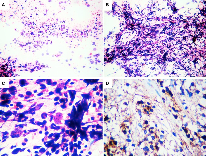
Fig. 1.
In the FNAC samples we observed a hemorrhagic background with neoplastic round and fibrillar cells with cytoplasmic processes, nuclear atypia, hyperchromatic nuclei with coarse chromatin, and some mitotic figures (a, HE 5×). These cells form clusters and tufts resembling glomeruli (b, HE 5×), Also observed were anaplastic figures, large pleomorphic cells with irregular nuclear outlines and prominent nucleoli (c, HE 40×), and immunoreactivity for GFAP (d, 40×). GFAP glial fibrillary acid protein

