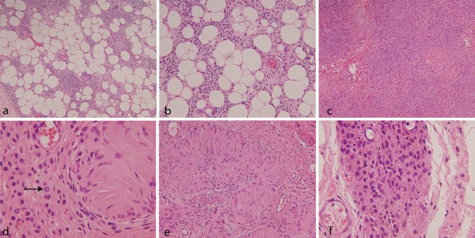
Fig. 2.
Showing HE stained sections of the lesion with diffuse infiltration by small, monotonous slightly spindled cells into the subcutaneous fat [a (×10), b (×20)]; adjacent solid area with a nested growth pattern, [c, (×10)]; presence of intra-nuclear inclusions [d, (×40)]; Wagner–Meissner bodies [e, (×20)]; and focal brown pigment [f, (×40)]

