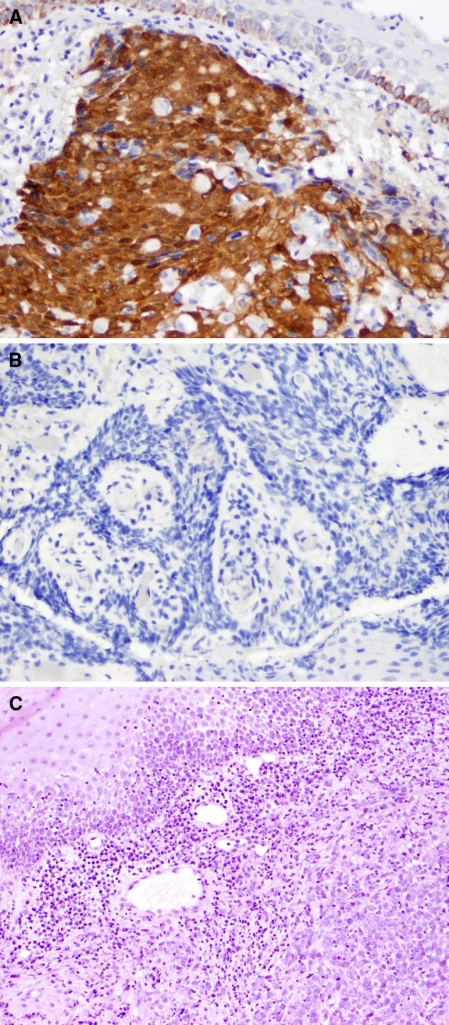
Fig. 3.

a p16INK4a positive OPSC (A) (40×); a p16INK4a negative OPSC b (40×). The histologic features (H & E stain) of a HPV-driven (p16INK4a positive and HPV positive) OPSC demonstrating non-keratinizing squamous cell carcinoma at lower right, surrounded by chronic inflammation with representative normal squamous epithelium seen at upper left c (20×)
