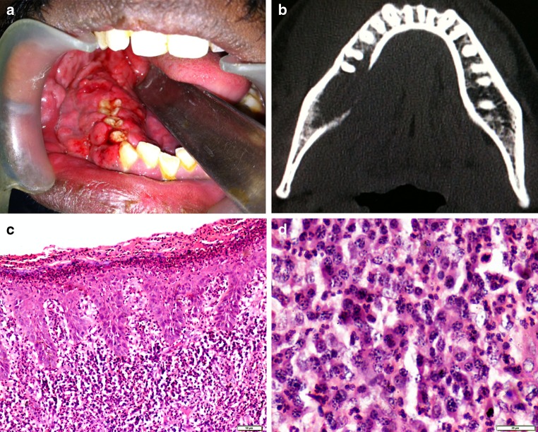
Fig. 1.
a Clinical photograph showing a soft tissue mass arising from extraction socket of right mandibular third molar. b Axial CT highlighting osteolytic areas with destruction of lingual cortical plate. c Highly cellular connective tissue stroma with surface erosions and psoriasiform epithelium (Haematoxylin and eosin, ×100). d Intense proliferation of plasma cells with scattered lymphocytes, neutrophils and eosinophils. (Haematoxylin and eosin, ×400)

