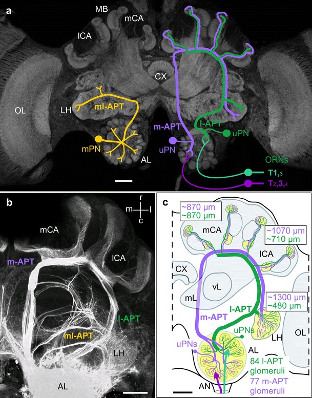
Fig. 1.

Anatomical features of parallel olfactory systems in the honeybee brain with special emphasis on a dual olfactory pathway from the antennal lobe (AL) to higher-order centers in the mushroom bodies (MB) and lateral horn (LH). a Schematic drawings of individual projection neurons (PN) from different antennal-lobe protocerebral tracts (APT) superimposed on a confocal image of the honeybee brain. Right brain half schematic drawings of a medial-tract uniglomerular PN (m-APT, uPN) and a lateral-tract PN (l-APT, uPN). Left brain half schematic drawing of a multiglomerular PN (mPN) that projects to the lateral horn (LH) only via the medio-lateral tract (ml-APT). The sensory input from olfactory receptor neurons (ORNs) to l- and m-APT associated glomeruli via four sensory-input tracts (T1-4) is schematically indicated on the right side. The size of the tract numbers depicts the dominance of different tracts in the two AL hemilobes. Adapted and modified with permission from Rössler and Zube (2011). b Projection view of an anterograde mass-fill of all APTs. Projection of the two major m- and l-APT from the AL to the medial and lateral MB calyces (mCA, lCA) of the MB and the LH. Three m- and l-APT (1–3) branch off the m-APT and innervate the lateral protocerebrum. Adapted and modified with permission from Kirschner et al. (2006). c Schematic overview of the dual olfactory pathway in the honeybee. ~84 glomeruli in the upper half of the AL are innervated by l-APT PNs that target the LH first and then the lCA and mCA. The m-APT originates from ~77 glomeruli in the lower half of the AL and projects to the mCA and lCA first before it targets the LH. The approximate distances of axonal trajectories via the m- and l-APT pathway to the three targets are indicated in green (l-APT) and magenta (m-APT). Adapted and modified with permission from Kirschner et al. (2006). AN antennal nerve, CX central complex, OL optic lobes, mL and vL medial and vertical lobes of the MB. Scale bars in a–c 100 μm
