
Fig. 3.
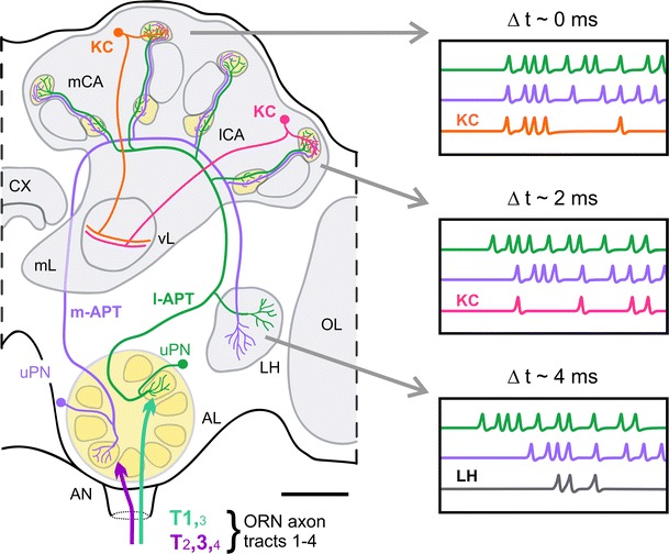
Model on a delay-line-like organization in the honeybee dual olfactory pathway based on anatomical and physiological findings (see text for details). Schematic drawings show individual medial and lateral antennal lobe protocerebral tract (m-, l-APT) uniglomerular projection neurons (uPNs). Schematic drawings of two individual Kenyon cells (KC) are included in the medial and lateral mushroom-body calyx (mCA, lCA) that receives convergent input from the two uPNs. The estimated differences in the delay of action potentials from m- and l-APT PNs at the mCA, lCA and lateral horn (LH) based on typical conduction velocities in the honeybee and differences in anatomical distances are indicated on top of the boxes on the right-hand side (see text for details). According to this model, differences in the delay of PN responses (Δt) at the mCA and lCA lead to hypothetical differences in coincident activation of the KC in the mCA and lCA, as well as in a hypothetical postsynaptic LH neuron that receives convergent input from PNs of both tracts. AN antennal nerve, AL antennal lobe, CX central complex, mL medial lobe of the MB, ORN olfactory receptor neuron, T1–4 sensory-input tracts 1–4, vL vertical lobe of the MB. Scale bar 100 μm