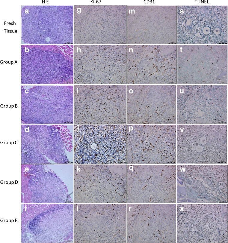
Fig. 2.
Representative photographs of grafted samples and ungrafted fresh tissue. a–f Hematoxylin and eosin stain. Original magnification × 100. g–l Ovarian section stained for Ki-67 expression. Gafted ovarian sections showed the intensive Ki-67 staining cells in the stroma. Original magnification × 400. m–r Ovarian section stained for CD31expression. Note the strong brown staining in the stroma of grafted ovarian section, indicating CD31 expression. Original magnification × 400. s–x Ovarian section stained by TUNEL assay. Original magnification × 400

