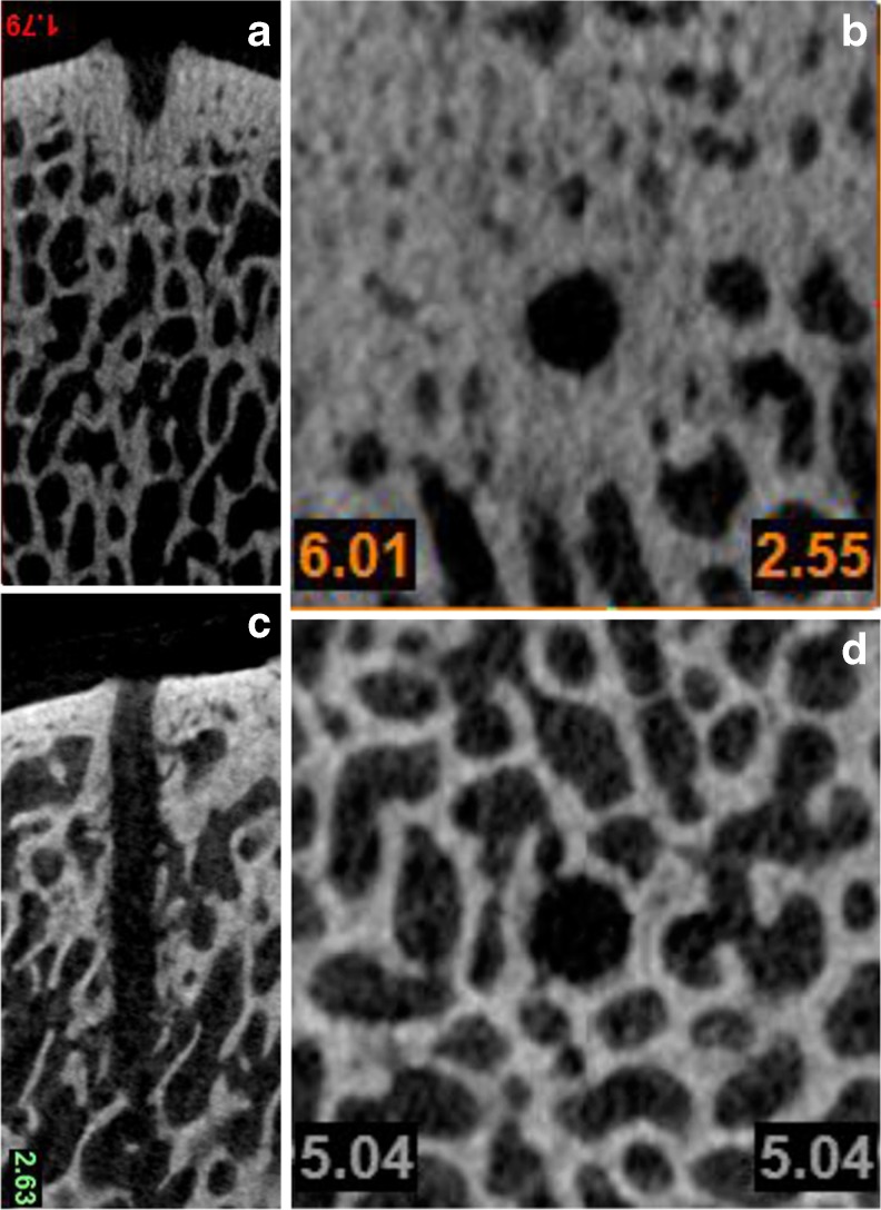
Fig. 5.
a–d Axial and sagittal microCT imaging. Adult bovine model courtesy of W.R. Walsh, Ph.D., N. Bertollo, Ph.D., D. Schaffner, M.D., R. Oliver, Ph.D., C. Christou BScVet. Surgical & Orthopaedic Research Laboratories (SORL), Prince of Wales Clinical School–The University of New South Wales, Sydney, Australia 2013. a Sagittal microCT following microfracture: dense bone compaction extending into cancellous bone with limited trabecular bone marrow channels. b Axial microCT depicting dense, compacted channel walls with well defined, highly regular channel walls. c Sagittal microCT following nanofracture: multiple open trabecular channels throughout the 9-mm deep subchondral bone perforation. d Axial microCT after nanofracture showing irregular wall outline and open trabecular channels. Trabecular bone structure appears to have normal thickness and density

