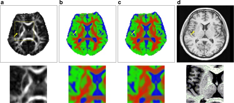
Fig. 5.
Images (top) at the external capsule level and magnifications (bottom) of the region. a The FA maps, b the estimated partial volume fraction maps obtained by the conventional method, c the estimated partial volume fraction maps obtained by proposed method, and d the structural images (T 1-weighted images). Yellow arrows point to the external capsules

