
Fig. 1.
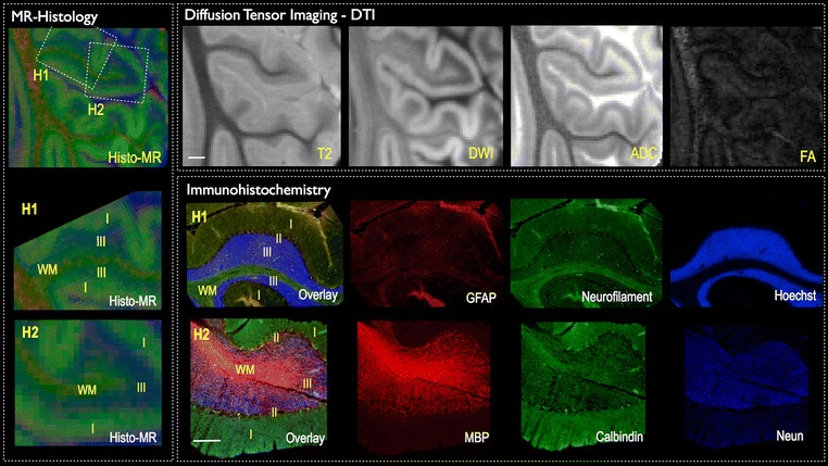
MR diffusion histology and immunohistochemistry. The cytoarchitectonic features of the cerebellar cortex are shown by diffusion imaging maps at 100 μm resolution. White matter (WM), granular layer (III), Purkinje cell layer (II) and molecular layer (I) can be discriminated across the different T2-weighted images, diffusion-weighted images (DWI), apparent diffusion coefficient (ADC) and fractional anisotropy (FA) contrasts (scale bar = 1 mm). A pseudo-colour visualisation of these maps is obtained by combining together FA (red), DWI (green) and ADC (blue) maps to mimic immunohistochemistry results and obtain a similar MR diffusion histology map. The comparison between MR diffusion histology and immunohistochemistry of the same region shows a good matching of features. Specifically, white matter tracts correspond to regions rich in myelin basic protein (MBP) and neurofilament, whereas a high cellular density, as indicated by the nuclear dye Hoechst, characterised the granular cell layer (III) (scale bar = 250 μm). These cells were predominantly neurons (NeuN + cells), whereas the Purkinje cell layer (II) was defined by calbindin + cells. However, the homogenous distribution of astrocytes (GFAP + cells) was not associated with MRI-detectable features