FIG. 5.
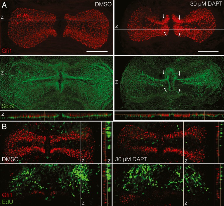
Hair cells appear to arise through transdifferentiation of support cells without proliferation. A In maximum intensity projections of P7 + 5 DIV cristae treated with 30 μm DAPT, the Sox9+ support cell layer (green) was disrupted near the eminentia cruciatum as compared to DMSO-treated controls where the Sox9 layer was continuous (arrows point to regions of increased hair cell density and decreased support cell density). This can also be seen in z projections through the sensory epithelium (at the white lines) where in controls the green support cell layer was continuous beneath the red hair cells, but in DAPT-treated cristae it was disrupted. This obvious disruption is not seen in adult explants. Scale bars 100 μm. B In P30 explants cultured for 5 DIV, hair cells did not take up EdU, despite the presence of EdU throughout the entire culture period. Cristae are shown in single slice views with labeling for Gfi1 (red) and EdU (green). z slice projections are shown to the right of the image indicating the location of the slice relative to the sensory epithelium in the z dimension. In both conditions, while many cells beneath the sensory epithelium were positive for EdU, no Gfi1+ hair cells had EdU labeling, as indicated by the lack of yellow cells.

