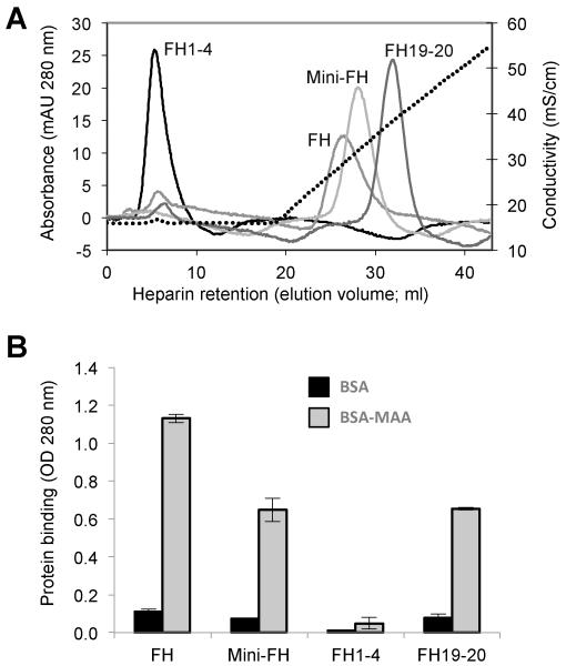
FIGURE 3.
Binding activity of mini-FH and FH-derived proteins to markers of self cells and oxidative stress. (A) Glycosaminoglycan binding as determined by heparin chromatography. Retention time during a NaCl gradient (0.15-0.5 M in phosphate buffer pH 7.4) signifies adhesion to heparin as model of polyanionic host surface pattern. (B) Recognition of oxidative damage markers. Binding of FH-derived proteins to surfaces coated with BSA or BSA modified with the lipid peroxidation product malondialdehyde-acetaldehyde (MAA-BSA) were analyzed by ELISA (detection with a polyclonal anti-FH Ab).

