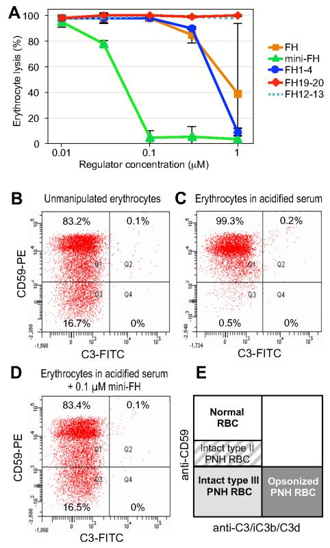
FIGURE 6.
Protection of PNH erythrocytes from AP-mediated lysis. (A) Erythrocytes were isolated from patient’s blood and incubated in acidified serum from ABO-matched healthy donors in the presence of various analytes, and lysis was determined after 24 h. (B-D) Prevention of C3 fragment deposition on PNH erythrocytes. Erythrocytes from patient’s blood (PNH patient #1) were exposed to AP activation by incubation in ABO-matched acidified serum. C3 deposition on PNH erythrocytes was assessed using an anti-C3 polyclonal antibody in combination with a counter staining with an anti-CD59 monoclonal antibody (35). Exposure to AP activation led to total disappearance of type III PNH erythrocytes (C) in comparison to freshly isolated erythrocytes (B). Mini-FH resulted in a full protection from lysis of type III PNH erythrocytes, without detectable deposition of C3 fragments on their surface (D). The scheme in panel E illustrates classification of erythrocytes as a function of surface molecules present (type II and type III PNH erythrocytes are defined according to the expression of CD59).

