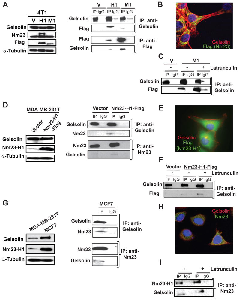
Figure 2. Gelsolin interacts with Nm23-H1 and -M1 in murine and human cell lines.
A. Left panel, western blot of lysates of 4T1 cells expressing either a vector (V), Flag-tagged Nm23-H1 (H1) or Flag-Nm23-M1 (M1). Right panel, co-immunoprecipitation of Gelsolin and Nm23 with either rabbit anti-Gelsolin or mouse anti-Flag antibody respectively. Either rabbit or mouse IgG was used as a control. B. Confocal microscopy image showed cytoplasmic co-immunoflorescence of Gelsolin (red) and Flag for Nm23-M1-Flag (green). C. Vector or Nm23-M1-Flag 4T1 cells were incubated 3 hours with either DMSO or 0.33μg/ml latrunculin B, and co-immunoprecipitation experiments performed to demonstrate Nm23/Gelsolin complex formation under actin-polymerization independent conditions. D. MDA-MB-231T human breast carcinoma cells were transfected with a vector or Flag-tagged Nm23-H1. Western blot (left) and co-immunoprecipitation (right) of Gelsolin and Flag-tagged Nm23-H1 from lysates was performed using mouse anti-Gelsolin and rabbit anti-Nm23 antibodies. E. Co-immunofluorescent staining was performed on MDA-MB-231T cells overexpressing Nm23-H1-Flag using mouse anti-Gelsolin red) and rabbit anti-Flag (green). F. MDA-MB-231T cells expressing either vector or Nm23-H1-Flag were incubated with either DMSO or 0.33μg/ml latrunculin B, and the lysates used for a co-immunoprecipitation experiment. G. Gelsolin and Nm23-H1 expression levels in MDA-MB-231T and MCF7 cell lines was analyzed by Western blot using mouse anti-Nm23-H1 and mouse anti-Gelsolin antibodies (left). The α-tubulin antibody was used as loading control. Protein lysate from MCF7 cells was used to pull down the either the endogenous Nm23-H1 with rabbit anti-Nm23-H1 antibody or Gelsolin with mouse anti-Gelsolin antibody. Western blots of the immunoprecipitates indicate complex formation between the endogenous Nm23-H1 and Gelsolin (right). H. Cytoplasmatic co-localization of Nm23-H1 and Gelsolin was identified in the immunoflorescent staining using mouse anti-Gelsolin (red) and rabbit anti-Nm23-H1 (green). I. Co-immunoprecipitation experiment was performed on MCF7 protein lysate after either DMSO or latrunculin B treatment of the cells in panel G.

