
Figure 1.
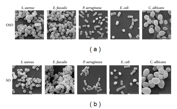
Scanning electron micrographs of the surface morphology of the cells after contact with either ozonated sesame oil (a) or sesame oil as control (b). Scale bars correspond to 2 μm, except for Candida albicans (5 μm). Arrowheads show small vesicles on cellular surface of Pseudomonas aeruginosa (see text for further details).