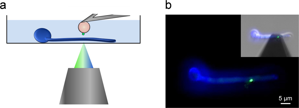
Fig. 1.
Single-cell force spectroscopy of bacterial-fungal interactions. (a) Schematic of the experimental set-up. (b) Using an integrated AFM-inverted optical microscope, the S. epidermidis probe (green) is approached towards a C. albicans hyphae (blue). The image was obtained using epifluorescence microscopy while the inset shows a merged phase/epifluorescence image.

