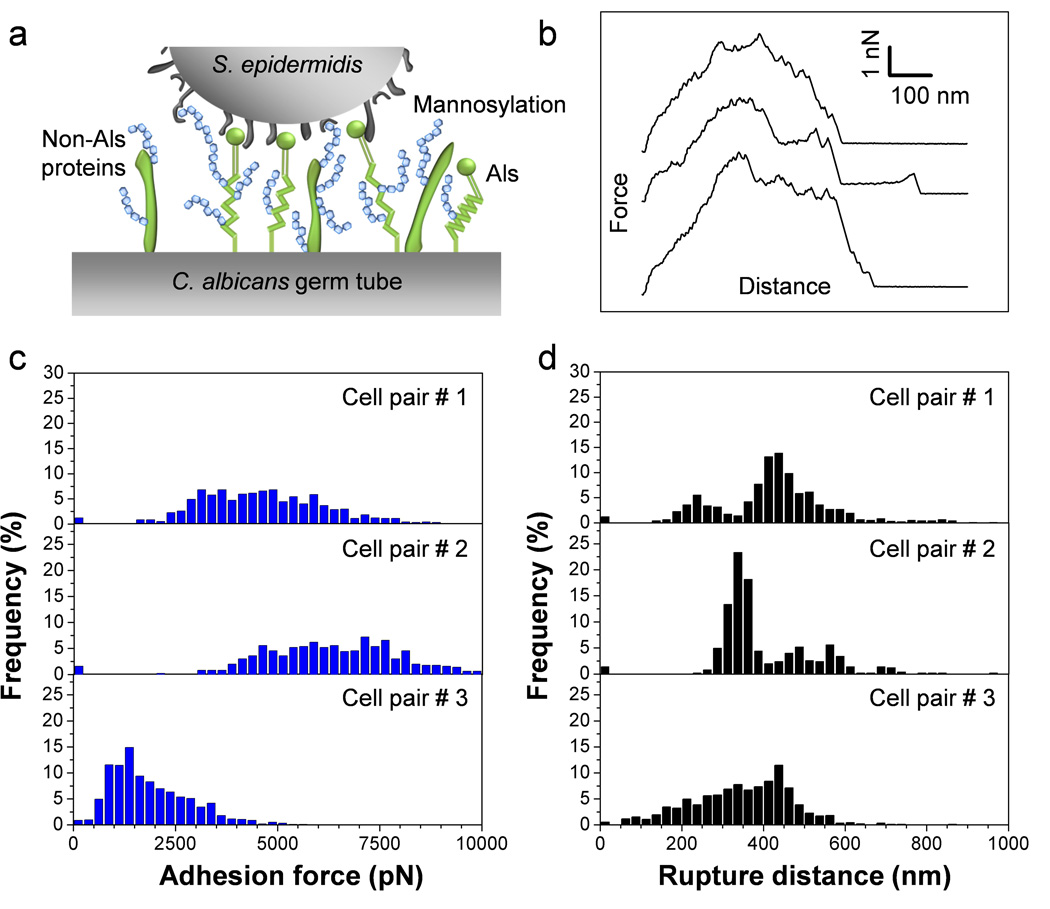
Fig. 2.
SCFS quantifies the adhesion forces between S. epidermidis and C. albicans germ tubes. (a) Key cell wall components that are involved in C. albicans surface interactions are cell-surface glycoproteins (in green) and mannose-rich glycoconjugates (in blue). (b) Typical force-distance curves recorded in Tris NaCl buffer between S. epidermidis and C. albicans hyphae. (c, d) Adhesion force (c) and rupture length (d) histograms obtained by recording force curves between 3 cell pairs from different cell cultures, and representative of a total of 7 cell pairs (n > 500 force-distances curves for each pair).

