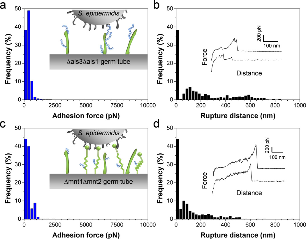
Fig. 5.
Als proteins and O-mannosylations on the C. albicans surface are required for bacterial adhesion. (a–d) Adhesion force (a, c) and rupture length (b, d) histograms, together with representative force curves, obtained by recording force curves in Tris NaCl buffer between a single S. epidermidis bacterium and a C. albicans germ tube from the mutant als3Δ/als3Δ als1Δ/als1Δ (Δals3Δals1) (a, b) or a C. albicans germ tube from the mutant mnt1Δ/mnt1Δ mnt2Δ/mnt2Δ (Δmnt1Δmnt2) (c, d). For each mutant, similar data were obtained in 3 independent experiments using 3 different cell pairs.

