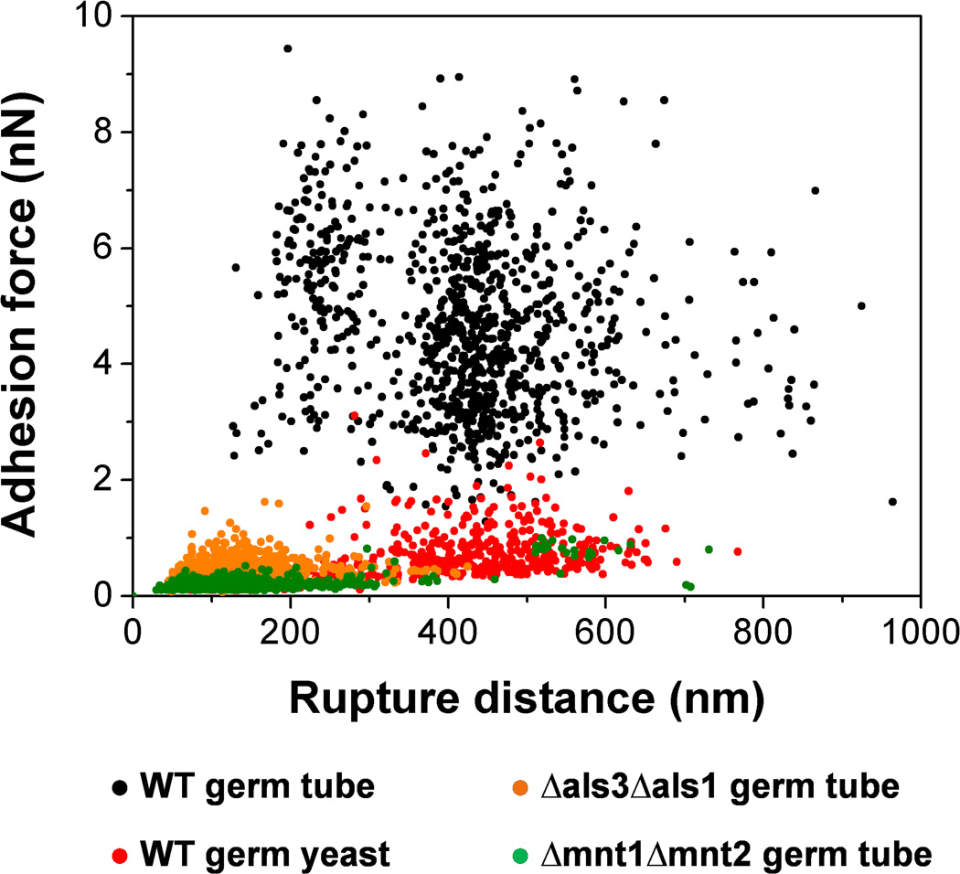
Fig. 6.
Role of cellular morphogenesis, Als proteins and O-mannosylations in S. epidermidis-C. albicans adhesion. Plots of the adhesion forces versus rupture distances measured between S. epidermidis and WT germ tubes (black symbols), WT germinated yeasts (red symbols), als3Δ/als3Δ als1Δ/als1Δ (Δals3Δals1) germ tubes (orange symbols) and mnt1Δ/mnt1Δ mnt2Δ/mnt2Δ (Δmnt1Δmnt2) germ tubes (green symbols). Strong co-adhesion is only observed on germ tubes and involves two types of highly adhesive and extended macromolecules, i.e. Als proteins and O-mannosylations.

