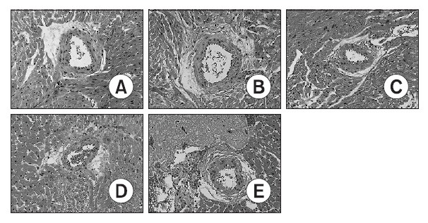
Fig. 3. Photomicrographs of hematoxylin/eosin stained heart sections from control-A and DOX-treated (3-B, 5-C, 7-D and 14-E) rats. DOX-treated hearts showed some inflammatory cells at periarterial area. Cytoplasmic vacuolization and nuclear degeneration were noted at heart tissues. Day 3-B, Day 5-C, Day 7-D and Day 14-E indicate the day after completion of a doxorubicin treatment period. Magnification ×200. A (0 day), B (3 days after doxorubicin treating), C (5 Day after doxorubicin treating), D (7 days after doxorubicin treating), E (14 days after doxorubicin treating).