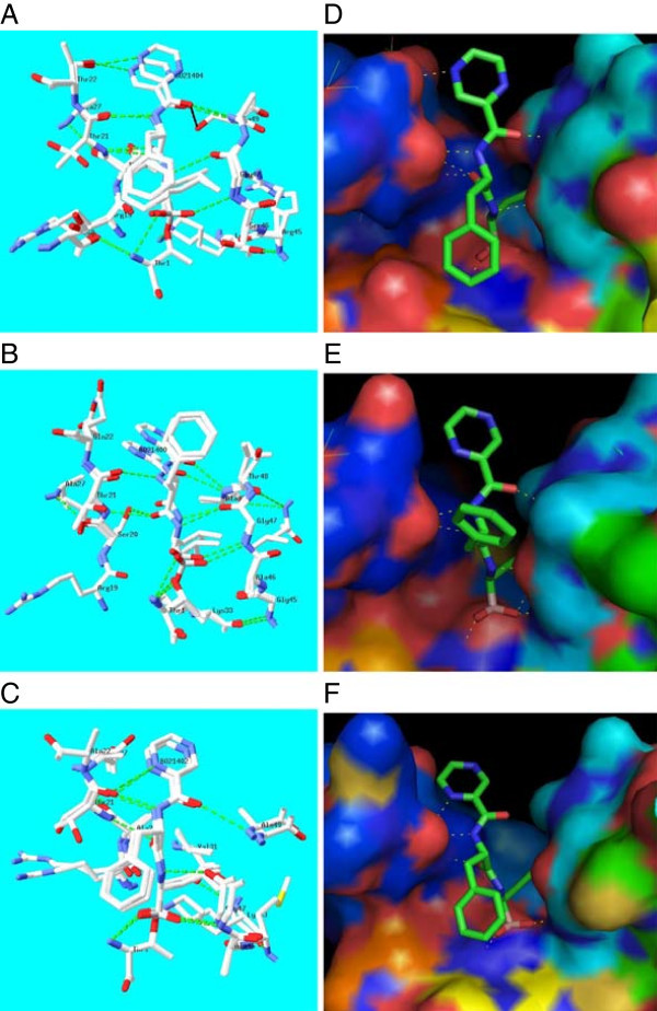
Figure 5.

Structural superposition of the yeast 20S proteasome - bortezomib inhibitor crystal structure complex [PDB code:2F16] and Pf 20S proteasome – bortezomib model showing inter-molecular hydrogen bond interactions and residues close to the inhibitor defined by a 4.0 Å cut-off value in (A) β1, (B) β2 and (C) β5 subunits. The electrostatic charge surface (red;electronegative, blue;electropositive) for the Pf 20S proteasome model with bound bortezomib for the above subunits are shown in figures (D), (E) and (F), respectively.
