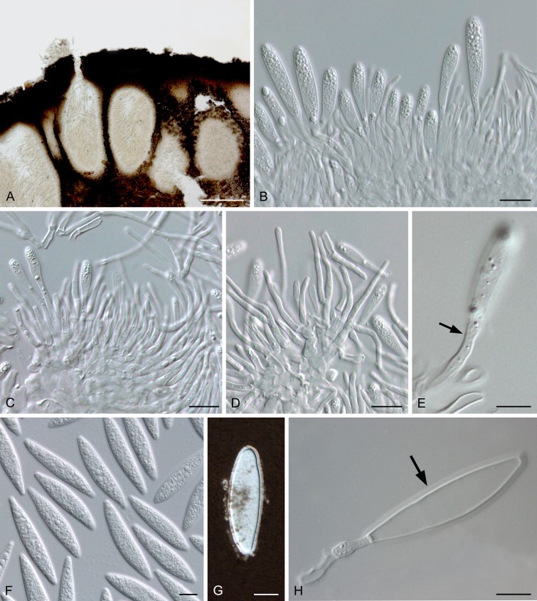
Fig. 6.
Melanopsaceae (Melanops tulasnei, LISE 95179). A. Section through conidiomata. B. Conidiogenous layers with developing conidia among paraphyses. C. Immature conidiogenous cells. D. Paraphyses. E. Conidiogenous cell with percurrent proliferations (arrowed). F. Conidia. G. Conidium in indian ink, revealing sheath. H. Conidium attached to conidiogenous cell with mucus sheath (arrowed). Scale bars: A = 200 μm, B-D, F, G = 10 μm, E, H = 5 μm (adapted from Phillips & Alves 2009).

