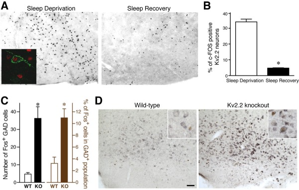
Figure 1.

Activity of Kv2.2-gamma-aminobutyric acidergic (GABAergic) neurons in the basal forebrain (BF). (A) The differential expression patterns of c-Fos in coronal sections that include the magnocellular preoptic area/horizontal limb of the diagonal band of Broca (MCPO/HDB) of the basal forebrain from sleep deprived and sleep recovered wild type (WT) mice. Inset: Double immunolabeling with anti-Kv2.2 (green) and anti–c-Fos (red) antibodies. (B) Quantitative analysis of three consecutive coronal brain sections (spanning 120 μm) containing MCPO/HDB from sleep deprived and sleep recovered WT mice. The percentages of c-Fos positive Kv2.2 neurons are shown (unpaired Student t-test; P = 0.0001; n = 4). (C) Augmented expression of c-Fos in GABAergic neurons in the BF of Kv2.2 knockout (KO) mice. The number (left) and percentage (right) of cells positive for both c-Fos protein and glutamic acid decarboxylase 67 (GAD67) messenger RNA (mRNA) in the MCPO are shown. *P < 0.01. (D) Double labeling of GAD67 mRNA (purple) and c-Fos protein (brown) in the MCPO. Scale bar, 100 μm. Inset: high-magnification images of neurons in the MCPO. Note that there are neurons double-positive for GAD67 mRNA and c-Fos protein in the KO section but not in the WT section.
