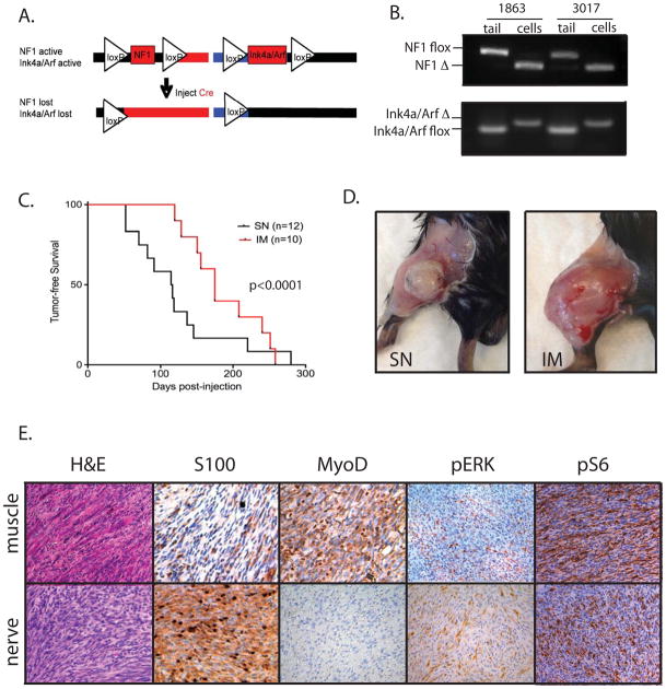
Figure 1. Characterization of a mouse model of NF1-deleted soft-tissue sarcoma.
A) Schematic of NF1 and Ink4a/Arf deletion following injection of an adenovirus expressing Cre recombinase. B) PCR demonstrating recombination of both NF1 and Ink4a/Arf floxed alleles in samples from paired tails and NF1-deleted sarcoma cell lines, derived from tumors generated by intramuscular injection of Ad-Cre. C) Kaplain-Meyer curve of tumor development based on site of orthotopic injection. The average time for tumor development is 4.1 months for mice injected in the sciatic nerve (SN) and 6.2 months for mice injected intramuscularly (IM). On right, photographs of NF1-deleted tumors generated from injections into the sciatic nerve (left) or muscle (right). D) Histopathology of IM and SN tumors. IM tumors resemble high-grade myogenic sarcomas and stain focally for MyoD1, but not S100. SN tumors resemble MPNSTs and stain focally for S100, but not for MyoD1. Both tumors are positive for pERK and pS6, indicating activity of the MAPK and mTOR pathways, respectively.

