
Fig. 5.
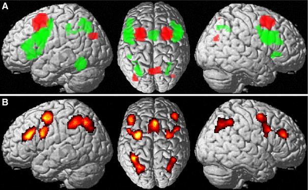
a Conjunction across task dependent (MACM) and task independent contrast analyses. Regions, which showed stronger connectivity with the posterior inferior frontal cortex are shown in green, while those regions, which showed stronger connectivity with the posterior superior frontal cortex are shown in red. b Conjunction across both approaches [task dependent (MACM) and independent (resting state) functional connectivity] and seeds (ventral and dorsal posterior inferior and posterior superior frontal cortex)