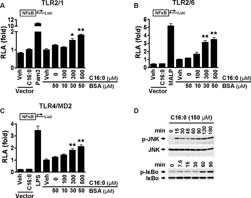
Figure 3. Palmitic acid induces NF-κB activation through TLR2 dimerized with TLR1 or TLR6 and through TLR4 and activates the downstream signaling pathways of pattern recognition receptors (PRRs).
(A-C) HEK293T cells were cultured in 10% FBS/DMEM medium and cotransfected with TLR2 and TLR1 (A), TLR2 and TLR6 (B), or TLR4 and MD2 (C) in addition to NF-κB-luciferase reporter and β–galactosidase expression vectors. After 24 hours the cells were serum-starved in 0.25% FBS/DMEM for 6 hours and then treated with C16:0-BSA for 12 hours. The cell lysates were assayed for luciferase and galactosidase activities. Values are expressed as RLA (relative luciferase activity). Controls: Pam3 (Pam3CSK4, TLR2/1 agonist, 10 ng/ml), MALP (MALP-2, TLR2/6 agonist, 10 ng/ml), LPS (TLR4 agonist, 50 ng/ml). pDisplay empty vector was used as a negative control. Data are expressed as mean ± SEM of three independent experiments. Significance was determined by unpaired, two-tailed, t test (*P < 0.05, **P < 0.01 significantly different from 50 µM BSA). (D) THP-1 cells were serum starved in 0.25% FBS-RPMI-1640 for 12 hours. Cells were treated with C16:0 (150 µM) for the indicated times and cell lysates were immunoblotted for phosphorylated JNK, total JNK, phosphorylated IκBα, and total IκBα.

