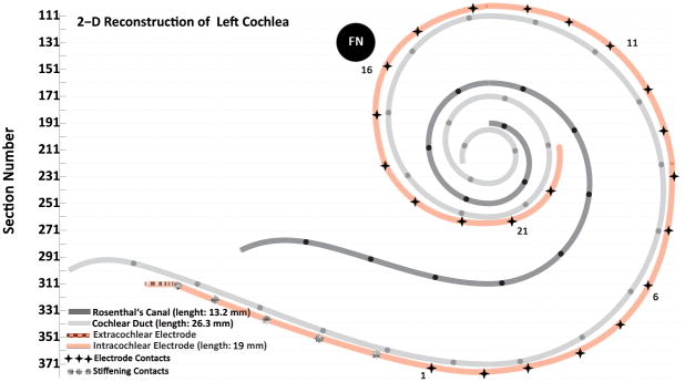
Figure 1.

Two-dimensional reconstruction of cochlear duct, Rosenthal’s canal and the electrode track of case 11. The electrode was fully inserted and stimulation of electrode contacts 14–18 caused facial nerve stimulation. The dark stars on the intracochlear electrode track mark the estimated position of the electrode contacts. The facial nerve (FN) was very close to the upper basal turn of cochlea (Figure 4). (The black circles on the line representing Rosenthal’s canal and on the line representing the cochlear duct are 1 mm apart.)
