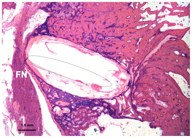
Figure 4.
Severe resorption and spongiosis in the bone between the facial nerve and upper basal turn of cochlea due to otosclerosis in case 11. In spite of severe thinning of the bone, there was still an otosclerotic bony septum separating the upper basal turn of cochlea from the facial nerve (FN). In this case the facial nerve was stimulated unintentionally by intracochlear stimulation. (The electrode was removed prior to sectioning the temporal bone. The location of the electrode track is shown by dotted line inside the cochlea.)

