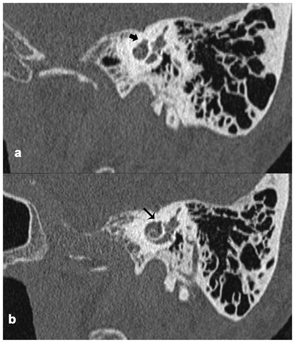
Figure 5.

Stenvers view of the temporal bone in CT scans of a living patient with FNS and otosclerosis. a. Involvement of the otic capsule by otosclerosis (thick black arrow). b. There was no bone separating the upper basal turn of cochlea and the facial nerve canal (thin black arrow) due to otosclerosis, suggesting the presence of dehiscence of the facial nerve.
