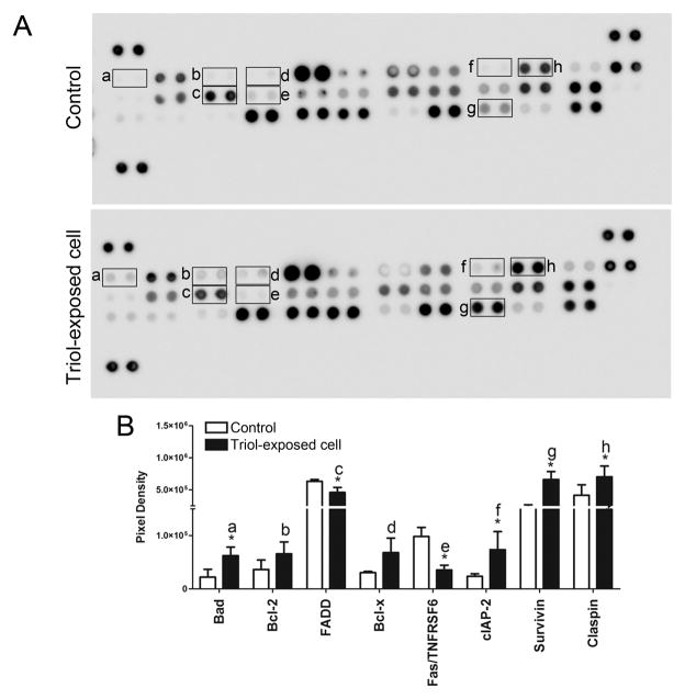
Fig. 4.
Activation of apoptosis-related proteins in Triol-exposed cells compares to MMNK-1 control cells. (A) Profiling of apoptosis-related proteins expression in control and Triol-exposed cells was done by using the apoptosis array. (B)The quantification of mean spot pixel densities were analyzed using ImageQuant™ from two individual experiments. * indicates P< 0.05.

