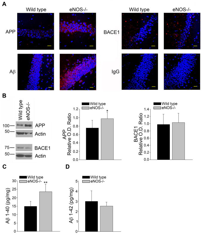
Figure 4.
APP and Aβ1-40 levels were increased in the hippocampus of LMA eNOS−/− mice. (A) Fixed tissue sections from the brains of wild type and eNOS−/− animals were immunolabeled with anti-APP, anti-BACE1 and anti-Aβ. Representative images of the hippocampus are shown. Magnification 40x; bar is representative of 20 μm. (B) Hippocampal tissue from LMA eNOS−/− and LMA wild type animals was Western blotted using anti-APP, anti-BACE1, and anti-Actin (loading control) antibodies. Representative image and densitometric analysis is shown. (C) Aβ1-40 and (D) Aβ1-42 levels from hippocampal lysates (200 μg total protein/sample) from LMA eNOS−/− and wild type control mice were analyzed via commercially available ELISA kits. Data are represented as mean ± SD (n=6 animals, *P<0.05, **P<0.01 compared to wild type control mice).

