
Figure 5.
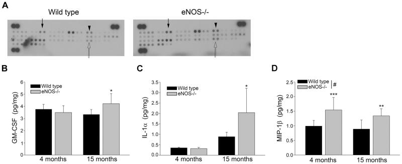
Levels of GM-CSF, IL-1α, and MIP-1β are increased in the brains of LMA eNOS−/− mice. (A) Brain lysates from LMA eNOS−/− and age-matched wild type control mice were analyzed via a commercially available cytokine array (300μg total protein loaded for each sample). A representative image is shown. Closed arrow depicts GM-CSF, arrowhead depicts IL-1α, and open arrow depicts MIP-1β. (B) GM-CSF (C) IL-1α, and (D) MIP-1β levels from 8 individual brain lysates (500 μg) from 4 month and 15 month old eNOS−/− and wild type control mice were analyzed via commercially available ELISA kits. Data are represented as mean ± SD (n=7–8 animals, *P<0.05, **P<0.01, and ***P<0.001 compared to age-matched wild type control mice; #P<0.05 based on genotype: 2-way ANOVA, followed by Tukey-Kramer post hoc tests for individual comparisons).