
Fig. 1.
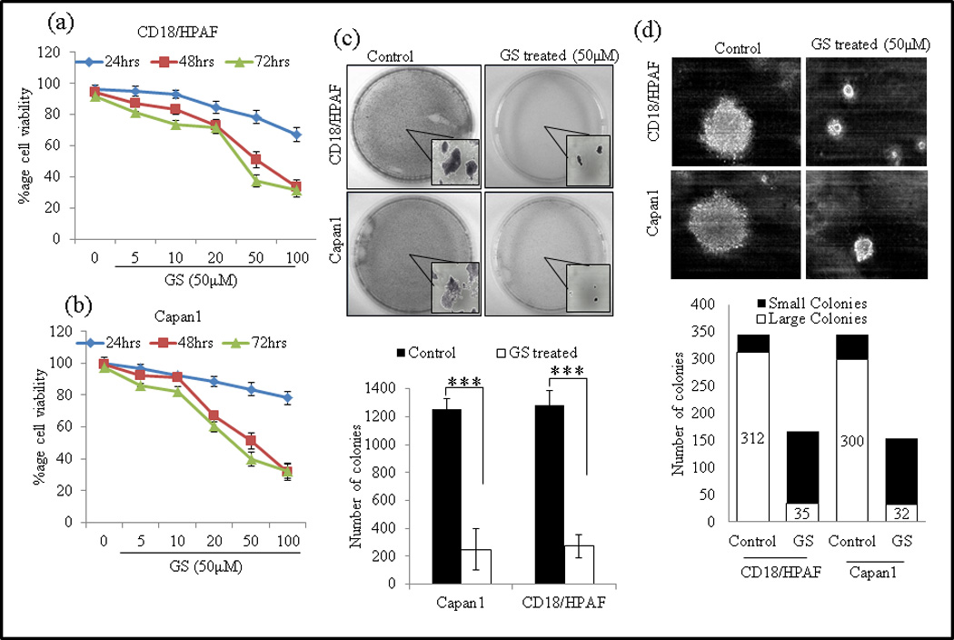
GS decreases proliferation of PC cell lines. PC cells Capan1 (a) and CD18/HPAF (b) were treated with different doses of GS for 24, 48 and 72 hrs and viable cell number was assayed by MTT assay. (c) GS reduces colony formation of PC cells. Capan1 and CD18/HPAF cells were incubated with GS (50µM) for 24 h and equal number of cells (1×10³) cells were seeded in triplicate in 10% DMEM in a 12-well plate, and allowed them to grow in 10% DMEM for 2 weeks. At the end of the incubation period, the colonies formed were washed with PBS, fixed in methanol and then stained with 0.1% Crystal violet in PBS. The number of colonies (per well) were counted with the automatic colony counting tool of the Quantity One Imaging software. The graphs represent the mean (±SE) number of colonies. (d) GS reduces anchorage-independent growth of PC cells in soft agar. GS treated or untreated Capan1 and CD18/HPAF/HPAF cells were incubated with GS (50µM) for 24 hrs and equal number of cells (2×10³) cells were seeded in triplicate with 0.35% agarose over the layer of 0.5% agarose and allowed them to grow 14 days. The colonies were counted and captured. The experiment were repeated twice (* p<0.05).