
Fig. 3.
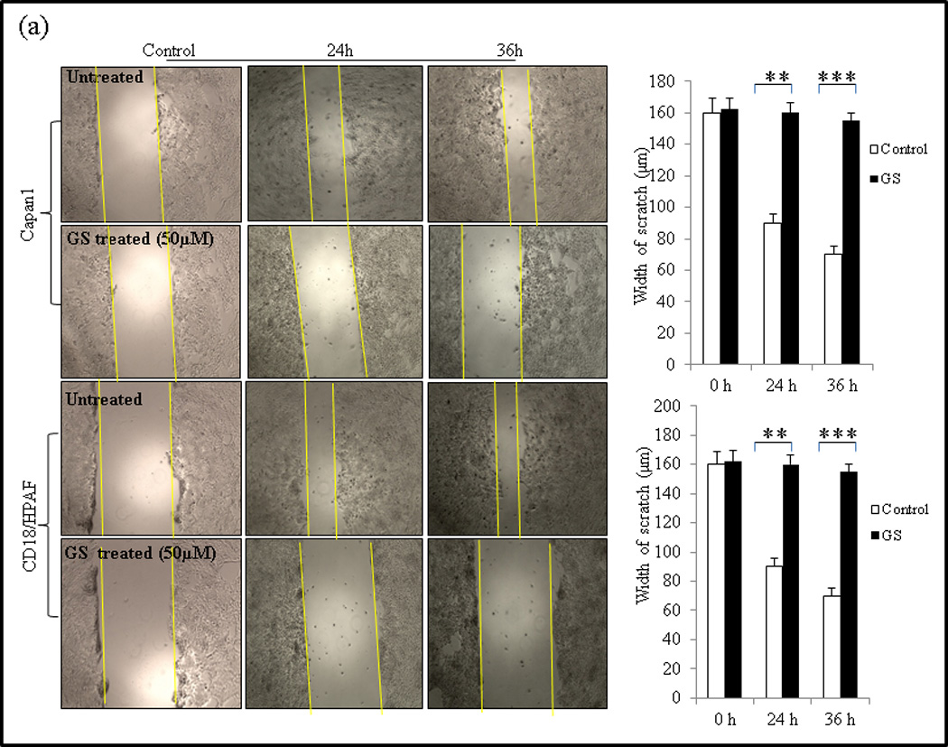
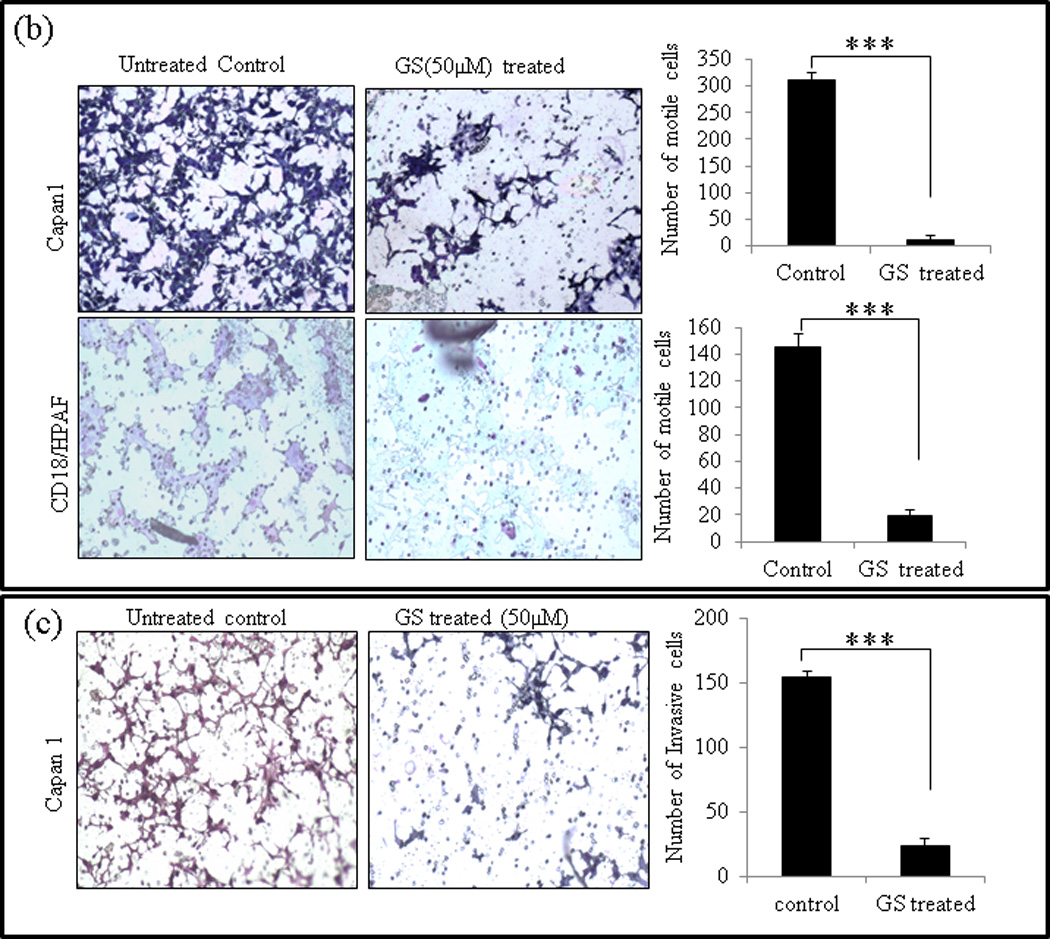
GS inhibits cell migration and invasion of PC cells. (a) Wound healing assay for evaluating the effect of GS on the motility of Capan1 and CD18/HPAF cells. Confluent monolayers of Capan1 and CD18/HPAF cells were scratched with a 200µl sterile pipette tip. Cells were carefully washed with 10% DMEM to remove the unattached cells and repair was monitored microscopically. Images were taken immediately (t = 0h) and after incubation with GS for 24 and 36 hrs. The representative photographs showed the same area at time zero and after 24 and 36h of incubation with or without GS. Columns, mean (n = 3); bars, SD. **, P<0.01, ***, P<0.001, statistically significant compared with the vehicle treated control. (b) Transwell migration assay showing the inhibitory effects of GS on Capan1 and CD18/HPAF cell migration. Equal number of GS treated PC cells (1 × 10⁵ cells) were seeded into the upper chamber in 1 ml of serum-free DMEM and the lower compartments were filled with 1 ml of DMEM containing 10% serum. After 24 hrs at 37°C, non-invading cells on the upper surface of the filter were wiped out with a cotton swab, and the invaded cells on the lower surface of the filter were fixed and stained using Diff Kit and counted under a light microscope (magnification, x100). (c) Matrigel assay indicating the inhibitory effects of GS on PC cell invasion. Capan1 cells were treated with GS (50µM) for 24 hrs and equal number of the cells (1 × 10⁶ cells) was seeded into the upper part of matrigel coated Transwell chamber as discussed above. The number of migrated cells through the 8µm pore of polyethylene terephtalate (PET) membrane was quantified in 10 random fields. Columns, mean of migrated and invasive cell number from three independent experiments; bars, SD. *, P<0.05, **, P<0.01, statistically significant.