Fig. 5.
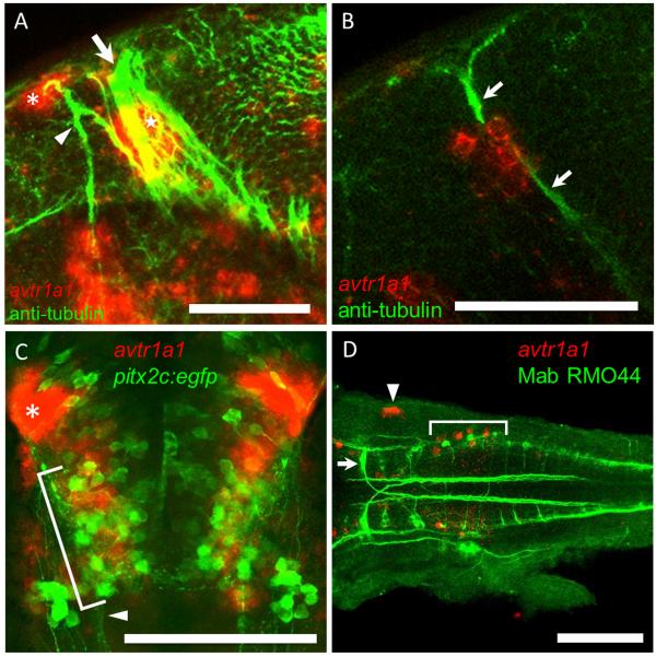
The epiphyseal and nucPC neurons but not the nucMLF nor T reticular neurons express avtr1a1 at approximately 48 hpf. (A) Lateral view (anterior left, dorsal up) of a z-stack of confocal images of a embryo double labeled for avtr1a1 and anti-acetylated tubulin (axons) showing that the cluster II avtr1a1+ cells at the lateral base of the epiphysis (red, asterisk) project axons into the DVDT (green, arrowhead) and the avtr1a1+ cluster III cells (red, star) are adjacent to the PC (arrow). The avtr1a1+ cells seen ventrally are the neurons in the ventral forebrain and near the forebrain/tegmentum boundary (I and IV). Scale: 50 um. (B) A single focal plane seen in a lateral view showing that the cluster III avtr1a1+ cells (red) appear to extend axons in the PC (green, arrows). Scale: 50 m. (C) Ventral perspective of pitx2c:egfp embryos labeled with a avtr1a1 riboprobe showing that the nucMLF neurons (green, bracket) do not express avtr1a1 (red). Arrowhead denotes the MLF; asterisk denotes the forebrain avtr1a1+ neurons. Scale: 100 m. (D) Dorsal perspective (anterior left) of the hindbrain of embryo double labeled for avtr1a1 riboprobe (red, bracket) and MAb RMO44 (green) showing that the posterior hindbrain avtr1a1+ cells are not the T reticular neurons. The arrowhead indicates avtr1a1+ cells in the otocyst. Scale: 100 m.

