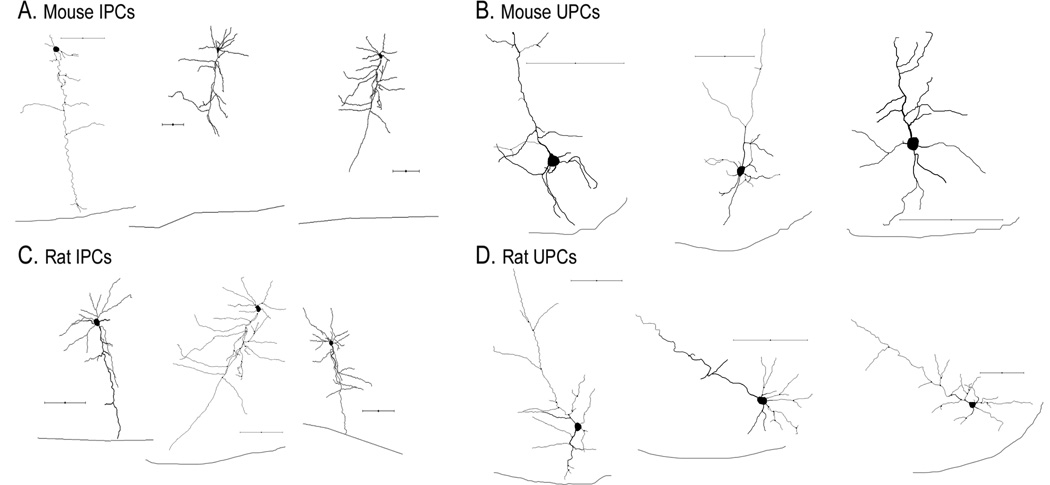
Figure 7.
Morphological reconstructions of inverted and upright pyramidal neurons following biocytin histochemistry. Representative reconstructions of mouse (A) and rat (C) IPCs. Representative reconstructions of mouse (B) and rat (D) UPCs. Black line below cells indicates layer VI-white matter border. All scalebars = 100µm.

