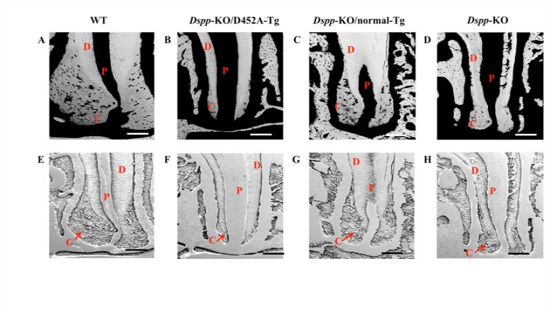
Figure 4. Failure to process DSPP into fragments leads to decreased cellular cementum deposition.
In these backscattered SEM (A-D) and resin-casted SEM (E-H) images, the dentin is denoted as “D”; pulp as “P” and cementum as “C”. In the backscattered SEM images, the black areas represent unmineralized or hypomineralized areas. Therefore, the pulpal space appears as black (denoted as P); Cementum (C) is less mineralized than Dentin (D) and appears darker than dentin. The Dspp-KO (D) and Dspp-KO/D452A-Tg mice (B) showed increased black areas periapically indicating decreased cellular cementum deposition compared to the WT (A) and Dspp-KO/normal-Tg mice (C). The resin-casted SEM gave a better visualization of the periapical region of cementum.. The WT (E) and Dspp-KO/normal-Tg mice (G) showed a thick layer of cementum deposition at the root apex whereas; Dspp-KO (H) and Dspp-KO/D452A-Tg mice (F) showed little or no cementum in the same region. Bar: 200 μm.

