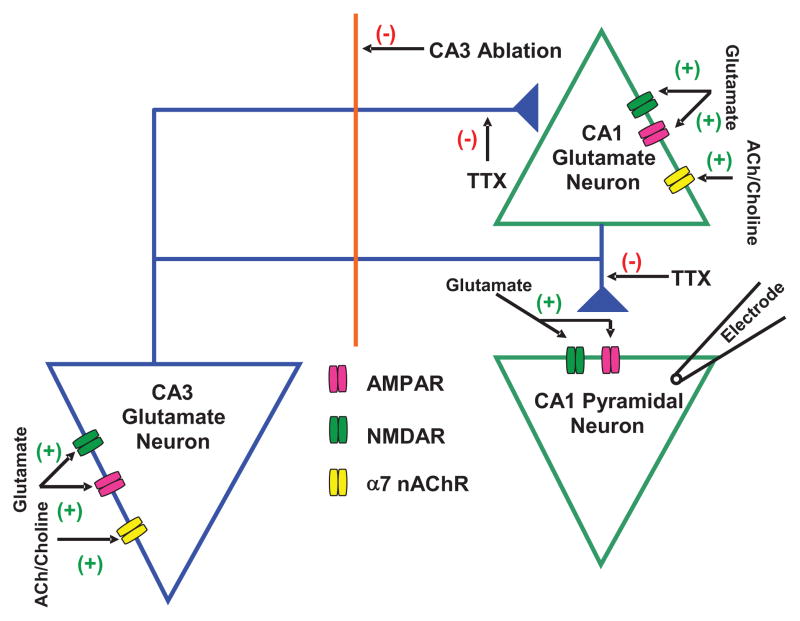
Figure 3. Schematic representation of glutamatergic transmission to CA1 pyramidal neuron.
Proposed model shows tonic regulation of glutamatergic input to CA1 pyramidal neurons by α7 nAChRs located on CA3 and CA1 pyramidal neurons. Spontaneous EPSCs were recorded from CA1 pyramidal neurons that received glutamate inputs from CA3 and CA1 pyramidal neurons. The α7 nAChR is reported to be present on CA3 and CA1 pyramidal neurons. The decrease in EPSC frequency by CA3 ablation suggests that CA3 pyramidal neurons regulate the excitability of CA1 pyramidal neurons. The magnitude of effect of MLA (10 nM) on EPSCs was larger in intact slices compared to that in CA3-ablated slices, which suggests that endogenous ACh or choline activates α7 nAChRs located on both CA3 and CA1 regions and controls glutamate activity in CA1 pyramidal neurons. (+) represents activation and (−) represents inhibition in the neurocircuitry.

