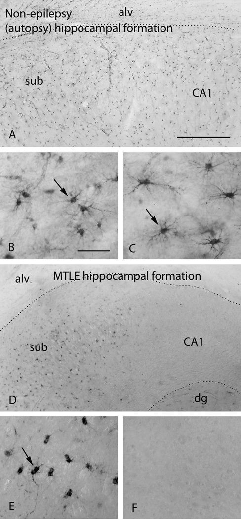
Figure 2.
Immunohistochemistry for GS reveals abundant labeling of astrocytes (arrows) in the subiculum (sub, A, B) and CA1 (A, C) of the hippocampal formation in a human autopsy subject without epilepsy. In contrast, GS is severely deficient in astrocytes in the hippocampal formation in a patient with mesial temporal lobe epilepsy [MTLE, (D–E)]. The deficiency is particularly pronounced in CA1 (D, F). Note that astrocytes in the subiculum of the MTLE hippocampus lack staining for GS in the most distal astrocyte processes (arrow in E) vs. the normal (autopsy) hippocampus (arrow in B). B and C are high power fields of the subiculum and CA1 respectively in A. E and F are high power fields of the subiculum and CA1 respectively in D. Abbreviations: alv, alveus; CA1, Cornu Ammonis subfield 1; dg, dentate gyrus; MTLE, mesial temporal lobe epilepsy. Scale bars: A=0.5 mm (same magnification as D); B=0.1 mm (same magnification as C, E, F). Reproduced from (Eid et al., 2004b), with permission from The Lancet/Elsevier.

