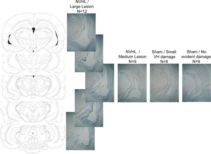
Figure 1.
Coronal sections through the ventral hippocampus from Paxinos and Watson (1998), and 5 vertically positioned photomicrographs of a typical “large” lesion in an NVHL group rat (n=12). Adjacent photomicrographs show typical damage at a roughly comparable AP plane (Bregma – 5.20 – 5.30 mm) in NVHL rats with a “medium” sized lesion (n=9), and in Sham group rats with little (n=6) or no damage noted to the NVHL (n=9).

