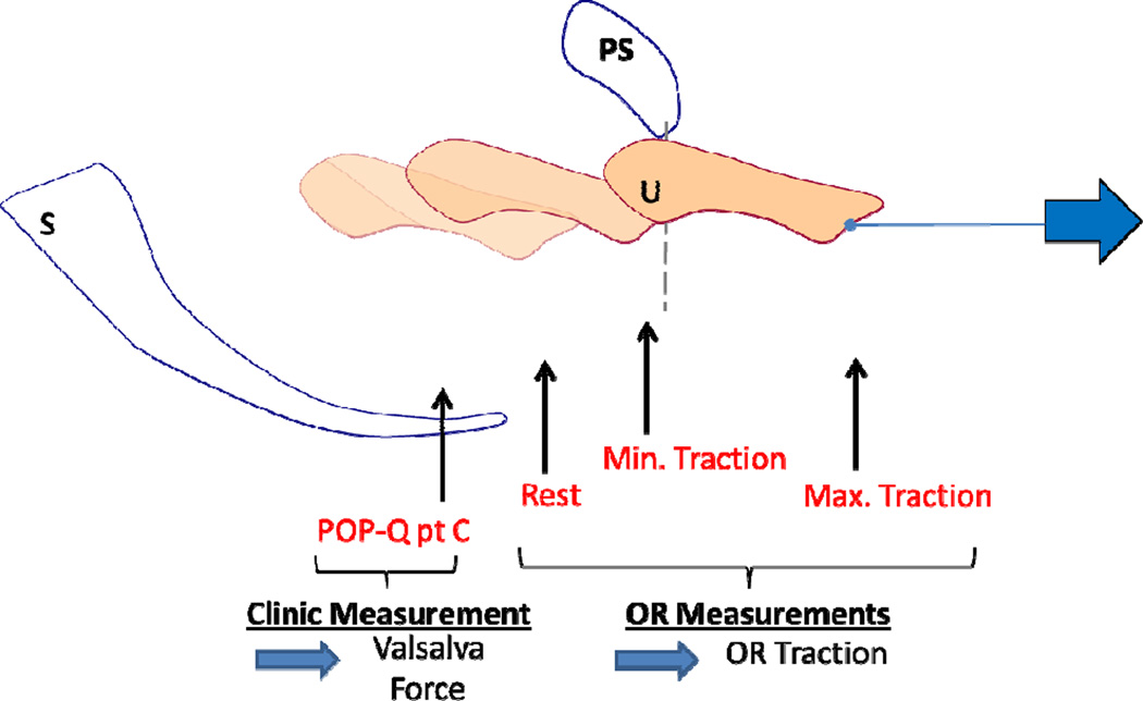
Figure 2. Cervix location measurements.
A schematic drawing of the measurements obtained during testing showing the uterus in four different locations in one subject (uterus (U), pubic symphysis (PS) and sacrum (S)). “POP-Q point C” indicates the location of the cervix measured in clinic at maximal Valsalva. Measurements “Rest”, “Min. Traction”, and “Max. Traction” indicate the cervix location at 0 force, minimal force (1.11N) and maximal force (17.8N) respectively. The latter measurements were taken in the operating room using the servoactuator.

