. 2013 Oct;37(4):389–400. doi: 10.5142/jgr.2013.37.389

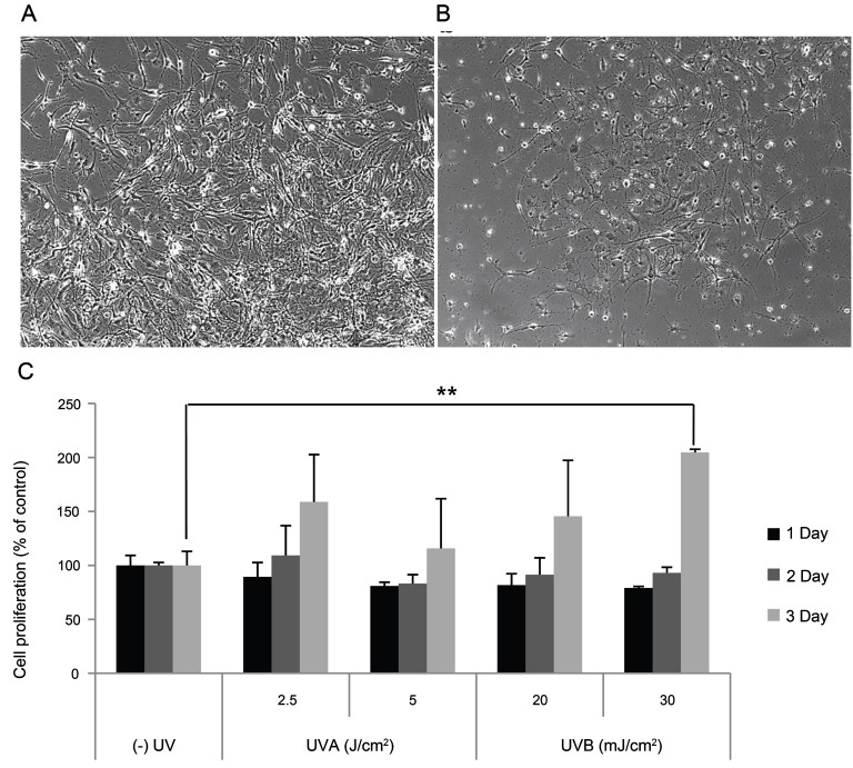
Fig. 4. Effect of conditioned media from UV-irradiated SP-1 keratinocytes on melan-A melanocyte proliferation. SP-1 keratinocytes were exposed to UVA (2.5, 5 J/cm2) or UVB (20, 30 mJ/cm2). After 24 h, the conditioned media was used to treat melan-A melanocytes. After 72 h, melan-A melanocyte proliferation was measured as described in the Materials and Methods. (A) UVB (30 mJ/cm2) irradiated conditioned media, (B) control, and (C) cell proliferation assay. Cells were observed under a phase-contrast microscope (Olympus CKX41; Olympus Optical, Tokyo, Japan) and images were photographed using a DMCe camera (INS Industry, Seoul, Korea) and the included DMC advanced software (×100). Data are shown as average±SEM. **p<0.01, compared with the control.

Fig. 4.