
Fig. 2A–B.
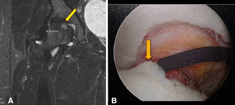
(A) A coronal STIR MR arthrography image shows a superior femoral head cartilage lesion (arrow) with underlying bone marrow edema (cursor), (B) correlating with intraoperative findings of a chondral flap lesion of the superior femoral head (arrow) with Grades II to III wear.