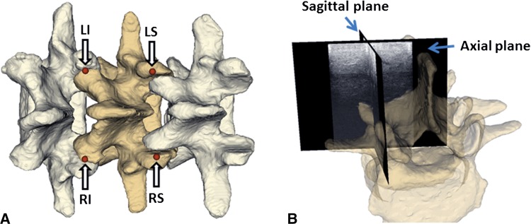
Fig. 2A–B.
(A) Arrows point to the four selected landmarks for vertebra registration. LI = left inferior; LS = left superior; RI = right inferior; RS = right superior. (B) The ultrasound snapshot image planes illustrate how to guide the sagittal plane to the facet joint area. The semitransparent vertebra overlaid on ultrasound snapshots is only for illustration and is not visible during actual landmark definition.

