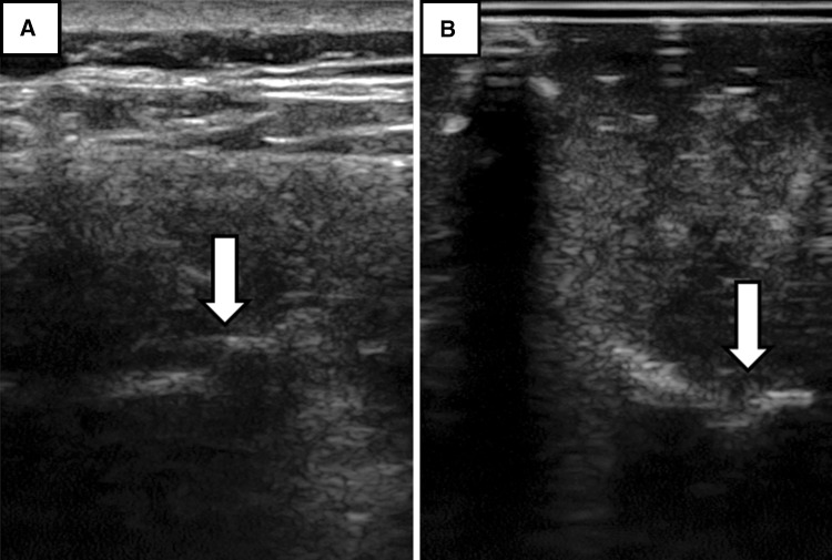
Fig. 3A–B.
(A) Example ultrasound images of the facet joint regions in the transverse plane in a human subject are shown. Arrows point to the facet joints. (B) Example ultrasound images of the facet joint regions in the transverse plane in a phantom model are shown. Arrows point to the facet joints.

