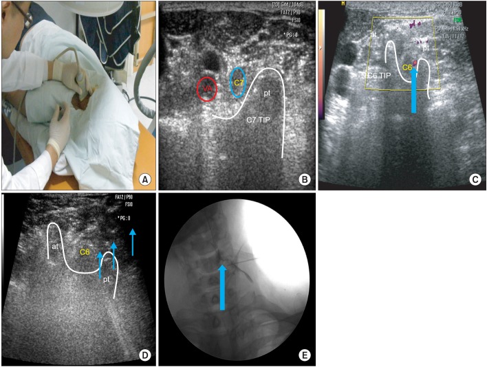
Fig. 1.
(A) Ultrasound-guided selective nerve root block (target nerve root C6). (B) This figure shows axial transverse ultrasound image of the C7 transverse process which has only 1 posterior tubercle. (C) Power Doppler image shows radicular arterial blood flows (arrowhead) of the C6 nerve root. (D) Needle (arrows) is placed on the dorsal surface of the C6 nerve root. (E) The targeted C6 nerve root is outlined by 1-2 mL contrast media (arrow). Contrast media spread to intraforaminal lesion (arrowhead). AT, anterior tubercle; PT, posterior tubercle.

