Figure 1.

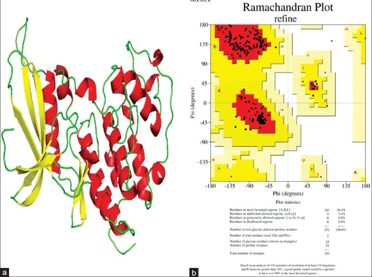
(a) Refined structure of the CDK4 protein, where helix is shown in red, yellow indicates beta sheets, and coil is shown in green color. (b) Ramachandran Plot of the protein after structural refinement

(a) Refined structure of the CDK4 protein, where helix is shown in red, yellow indicates beta sheets, and coil is shown in green color. (b) Ramachandran Plot of the protein after structural refinement